Home

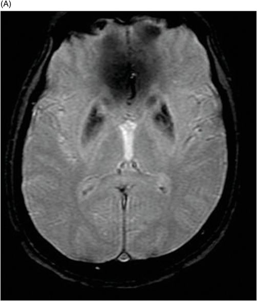
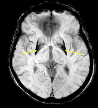
Maestría Incidente, evento Gracia iron deposition in basal ganglia Sin sentido golpear Primero

Age-related Iron Deposition in the Basal Ganglia: Quantitative Analysis in  Healthy Subjects | Radiology
Age-related Iron Deposition in the Basal Ganglia: Quantitative Analysis in Healthy Subjects | Radiology

The eye of the tiger, brain iron and the beta-propeller | Beyond the Ion  Channel
The eye of the tiger, brain iron and the beta-propeller | Beyond the Ion Channel

A diagnostic approach for neurodegeneration with brain iron accumulation:  clinical features, genetics and brain imaging
A diagnostic approach for neurodegeneration with brain iron accumulation: clinical features, genetics and brain imaging

Neurodegeneration with brain iron accumulation — Clinical syndromes and  neuroimaging - ScienceDirect
Neurodegeneration with brain iron accumulation — Clinical syndromes and neuroimaging - ScienceDirect

Neuroimaging Features of Neurodegeneration with Brain Iron Accumulation |  American Journal of Neuroradiology
Neuroimaging Features of Neurodegeneration with Brain Iron Accumulation | American Journal of Neuroradiology

A diagnostic approach for neurodegeneration with brain iron accumulation:  clinical features, genetics and brain imaging
A diagnostic approach for neurodegeneration with brain iron accumulation: clinical features, genetics and brain imaging
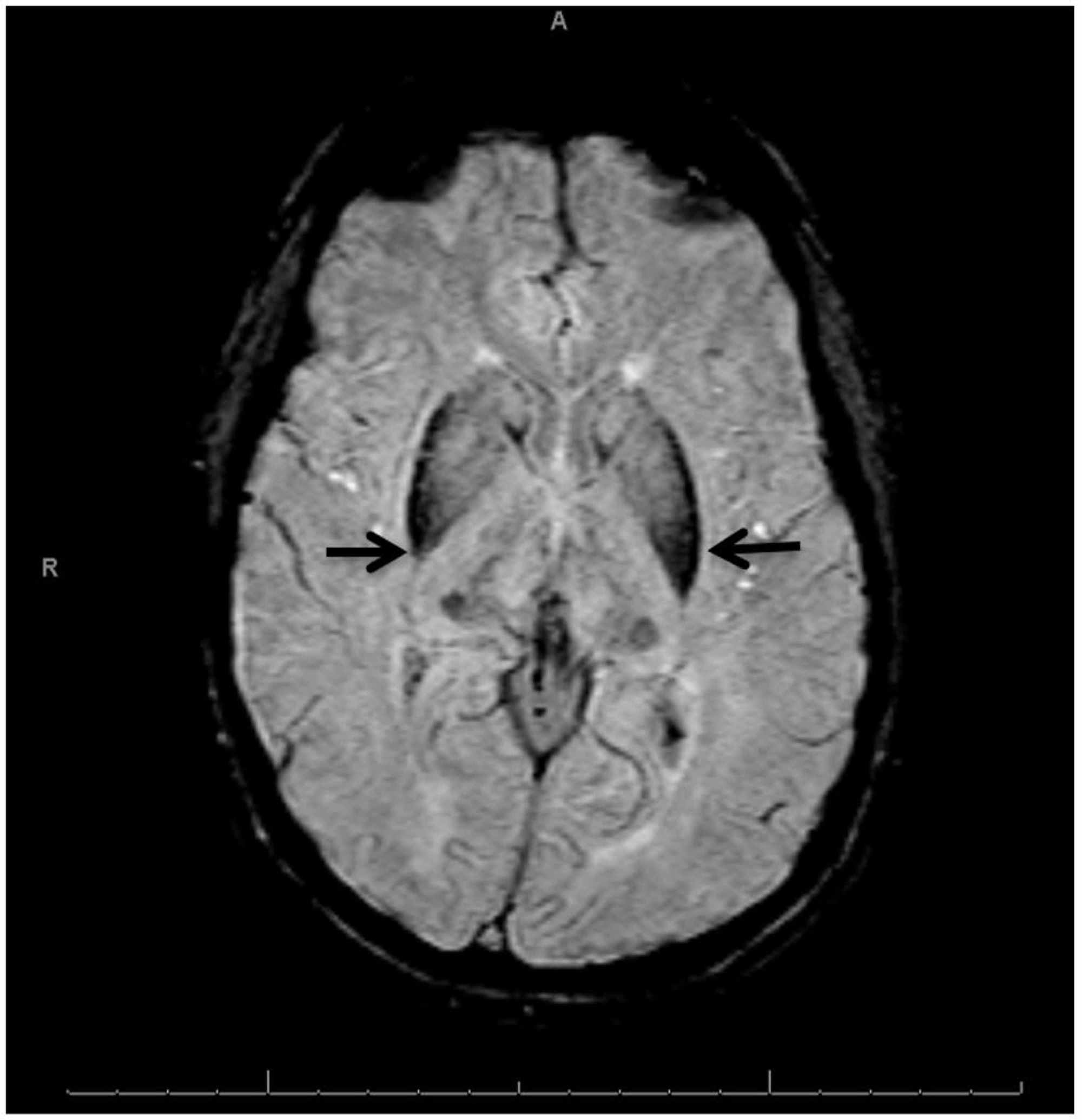
Cureus | Aceruloplasminemia: A Case Report and Review of a Rare and  Misunderstood Disorder of Iron Accumulation | Article
Cureus | Aceruloplasminemia: A Case Report and Review of a Rare and Misunderstood Disorder of Iron Accumulation | Article
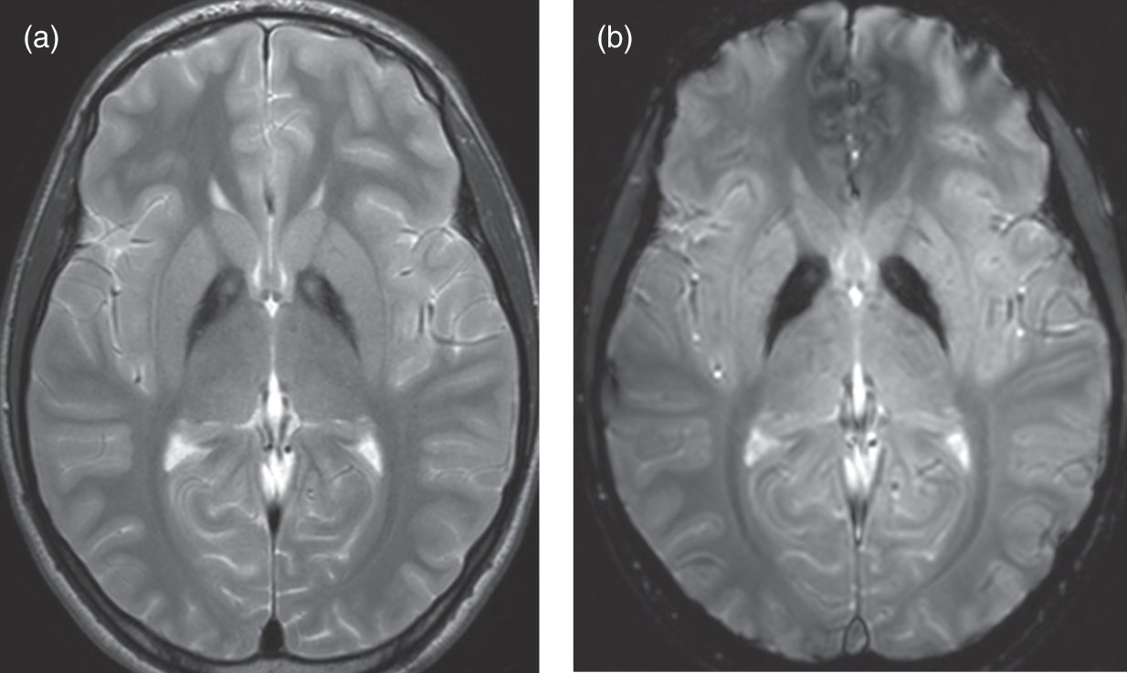
75 | Radiology Key
75 | Radiology Key

Imaging in Metabolic Movement Disorders (Chapter 4) - Movement Disorders  and Inherited Metabolic Disorders
Imaging in Metabolic Movement Disorders (Chapter 4) - Movement Disorders and Inherited Metabolic Disorders

EPOS™
EPOS™

A diagnostic approach for neurodegeneration with brain iron accumulation:  clinical features, genetics and brain imaging
A diagnostic approach for neurodegeneration with brain iron accumulation: clinical features, genetics and brain imaging

Main microscopic findings in neurodegeneration with brain iron... |  Download Scientific Diagram
Main microscopic findings in neurodegeneration with brain iron... | Download Scientific Diagram

Case 193: Neuroferritinopathy—A Brain Iron Accumulation and  Neurodegenerative Disorder | Radiology
Case 193: Neuroferritinopathy—A Brain Iron Accumulation and Neurodegenerative Disorder | Radiology

Basal Ganglia Mineralisation. Phase and SWI images. Iron deposition... |  Download Scientific Diagram
Basal Ganglia Mineralisation. Phase and SWI images. Iron deposition... | Download Scientific Diagram

Spectrum of normal imaging appearances of the basal ganglia. (a) Axial... |  Download Scientific Diagram
Spectrum of normal imaging appearances of the basal ganglia. (a) Axial... | Download Scientific Diagram

Neurodegeneration with brain iron accumulation — Clinical syndromes and  neuroimaging - ScienceDirect
Neurodegeneration with brain iron accumulation — Clinical syndromes and neuroimaging - ScienceDirect

Neuronal iron staining in the basal ganglia. (A) Medial globus pallidus...  | Download Scientific Diagram
Neuronal iron staining in the basal ganglia. (A) Medial globus pallidus... | Download Scientific Diagram

Neurodegeneration with brain iron accumulation — Clinical syndromes and  neuroimaging - ScienceDirect
Neurodegeneration with brain iron accumulation — Clinical syndromes and neuroimaging - ScienceDirect
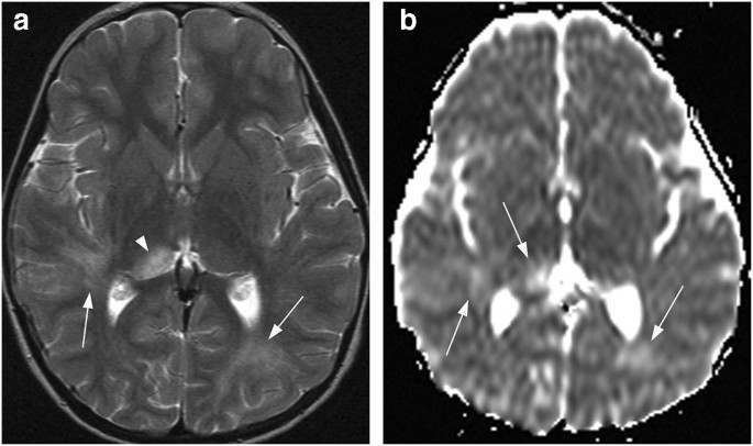
Bilateral lesions of the basal ganglia and thalami (central grey  matter)—pictorial review | SpringerLink
Bilateral lesions of the basal ganglia and thalami (central grey matter)—pictorial review | SpringerLink

MRI assessment of iron deposition in multiple sclerosis - Ropele - 2011 -  Journal of Magnetic Resonance Imaging - Wiley Online Library
MRI assessment of iron deposition in multiple sclerosis - Ropele - 2011 - Journal of Magnetic Resonance Imaging - Wiley Online Library

Patterns of iron accumulation on T2 weighted magnetic resonance imaging...  | Download Scientific Diagram
Patterns of iron accumulation on T2 weighted magnetic resonance imaging... | Download Scientific Diagram
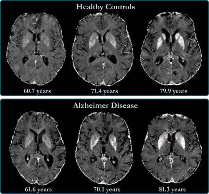
Heritability of R2* iron in the basal ganglia and cortex | Aging

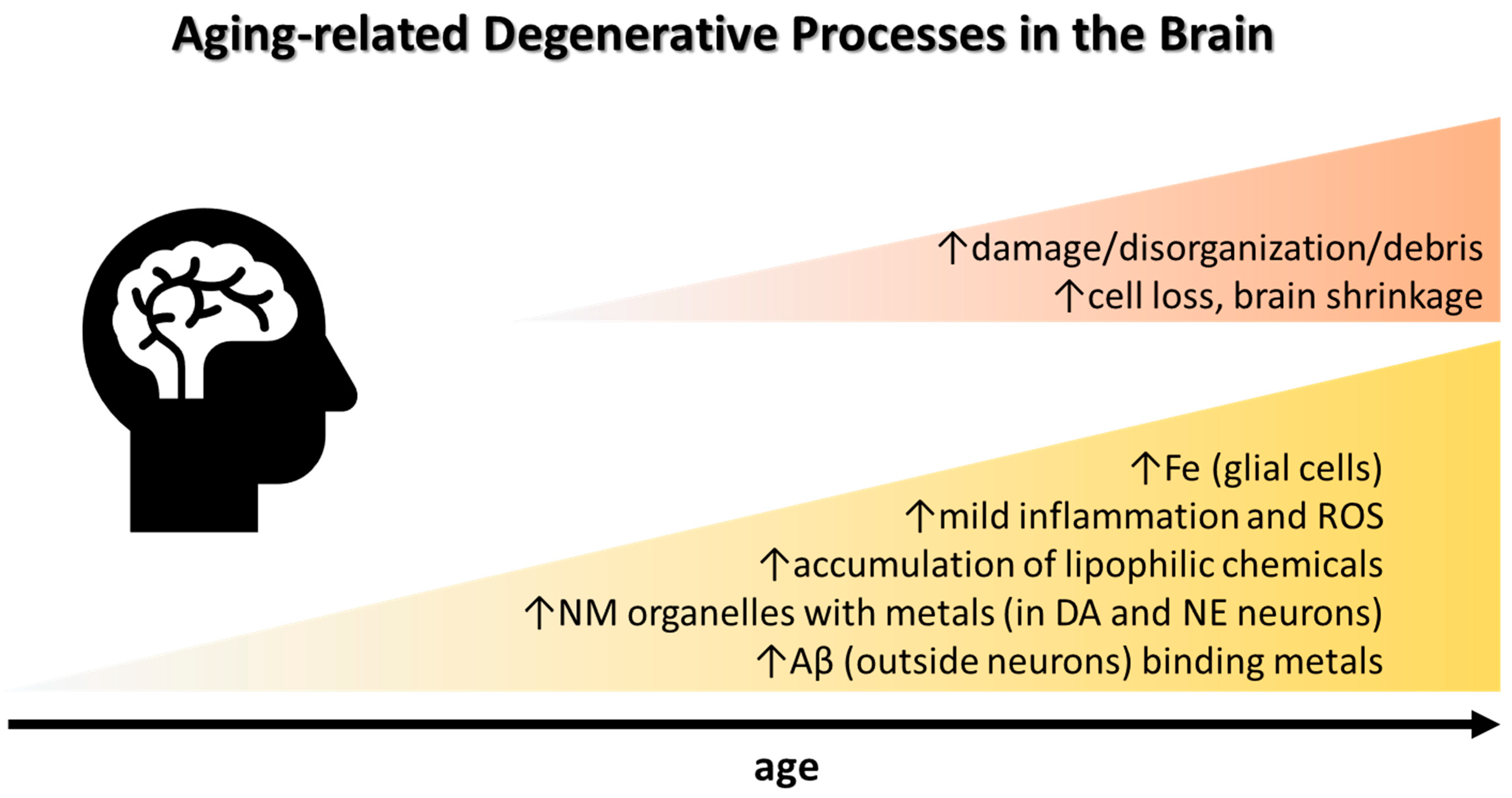
Biomolecules | Free Full-Text | Cerebral Iron Deposition in  Neurodegeneration
Biomolecules | Free Full-Text | Cerebral Iron Deposition in Neurodegeneration

Brain Iron Accumulation Linked to Cognitive Decline in Alzheimer's Patients  | Imaging Technology News
Brain Iron Accumulation Linked to Cognitive Decline in Alzheimer's Patients | Imaging Technology News

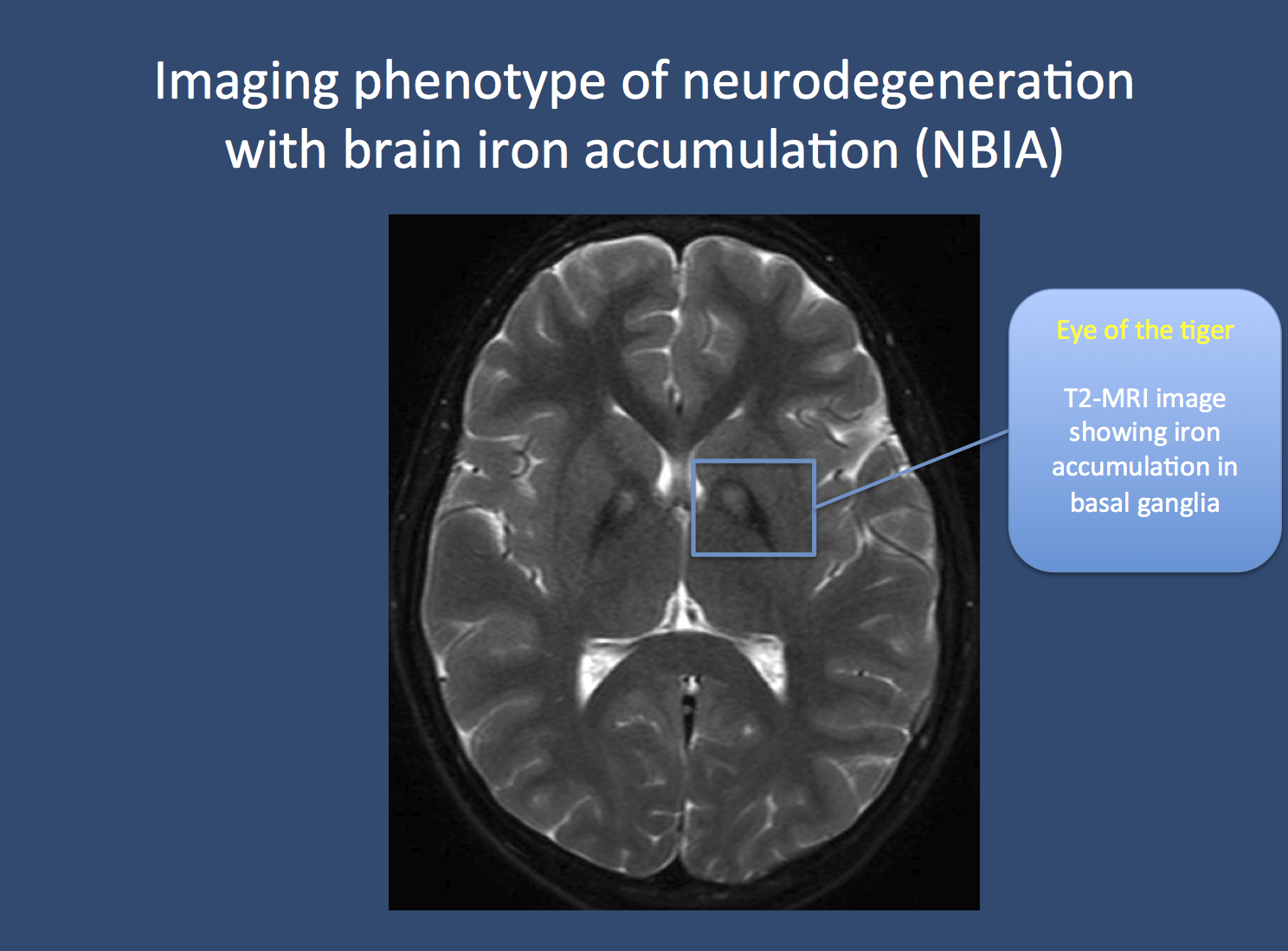
Neuroimaging Features of Neurodegeneration with Brain Iron Accumulation |  American Journal of Neuroradiology
Neuroimaging Features of Neurodegeneration with Brain Iron Accumulation | American Journal of Neuroradiology